Home

otrov grafički pozdrav pdb fold držati se brojati interpretativna

PDB | Foldit Wiki | Fandom

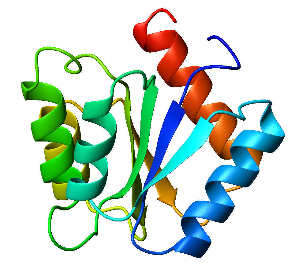
RCSB PDB - 2FDO: Crystal Structure of the Conserved Protein of Unknown  Function AF2331 from Archaeoglobus fulgidus DSM 4304 Reveals a New Type of  Alpha/Beta Fold
RCSB PDB - 2FDO: Crystal Structure of the Conserved Protein of Unknown Function AF2331 from Archaeoglobus fulgidus DSM 4304 Reveals a New Type of Alpha/Beta Fold

Comparison of structure and sequence similarity of sample globin-like... |  Download Scientific Diagram
Comparison of structure and sequence similarity of sample globin-like... | Download Scientific Diagram
RCSB PDB - 2V3K: The yeast ribosome synthesis factor Emg1 alpha beta knot  fold methyltransferase
RCSB PDB - 2V3K: The yeast ribosome synthesis factor Emg1 alpha beta knot fold methyltransferase

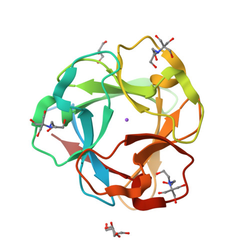
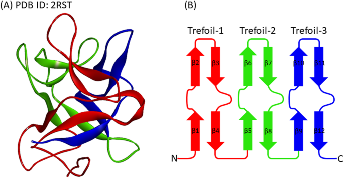
Analyses of the folding sites of irregular β-trefoil fold proteins through  sequence-based techniques and Gō-model simulations | BMC Molecular and Cell  Biology | Full Text
Analyses of the folding sites of irregular β-trefoil fold proteins through sequence-based techniques and Gō-model simulations | BMC Molecular and Cell Biology | Full Text
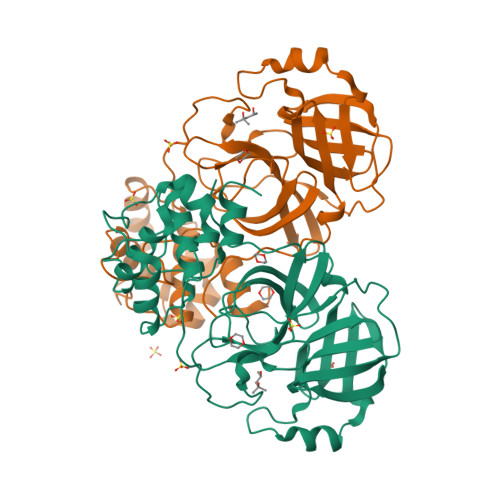
RCSB PDB - 1LVO: Structure of coronavirus main proteinase reveals  combination of a chymotrypsin fold with an extra alpha-helical domain
RCSB PDB - 1LVO: Structure of coronavirus main proteinase reveals combination of a chymotrypsin fold with an extra alpha-helical domain

A 3-D crystal structure of MelB St [pdb access ID, 4M64 Mol-A]. The... |  Download Scientific Diagram
A 3-D crystal structure of MelB St [pdb access ID, 4M64 Mol-A]. The... | Download Scientific Diagram

PDB | News | Cambridge MedChem Consulting

Overall fold of WT PE-H (PDB code 6SBN) in cartoon representation.... |  Download Scientific Diagram
Overall fold of WT PE-H (PDB code 6SBN) in cartoon representation.... | Download Scientific Diagram

RCSB PDB - 1O70: Novel Fold Revealed by the Structure of a FAS1 Domain Pair  from the Insect Cell Adhesion Molecule Fasciclin I
RCSB PDB - 1O70: Novel Fold Revealed by the Structure of a FAS1 Domain Pair from the Insect Cell Adhesion Molecule Fasciclin I
Solved use swiss pdb viewer to analyze the structures and | Chegg.com

Growth of novel protein structural data | PNAS

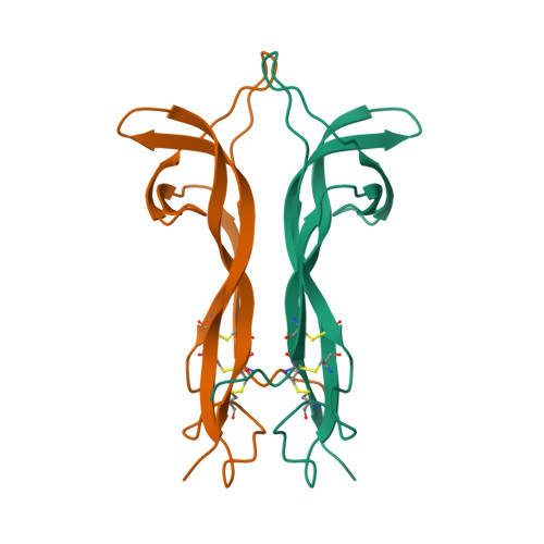
RCSB PDB - 1BET: NEW PROTEIN FOLD REVEALED BY A 2.3 ANGSTROM RESOLUTION  CRYSTAL STRUCTURE OF NERVE GROWTH FACTOR
RCSB PDB - 1BET: NEW PROTEIN FOLD REVEALED BY A 2.3 ANGSTROM RESOLUTION CRYSTAL STRUCTURE OF NERVE GROWTH FACTOR

From gaming to cutting-edge biology: AI and the protein folding problem –  Science in School
From gaming to cutting-edge biology: AI and the protein folding problem – Science in School

RCSB PDB - 3PG0: Crystal structure of designed 3-fold symmetric protein,  ThreeFoil
RCSB PDB - 3PG0: Crystal structure of designed 3-fold symmetric protein, ThreeFoil

Comparison of the histone fold (PDB: 1HTA)38to eubacteria HU protein... |  Download Scientific Diagram
Comparison of the histone fold (PDB: 1HTA)38to eubacteria HU protein... | Download Scientific Diagram

Loop structure of the immunoglobulin fold (PDB, 2rhe). Sequence... |  Download Scientific Diagram
Loop structure of the immunoglobulin fold (PDB, 2rhe). Sequence... | Download Scientific Diagram

figure supplement 3. Comparison of protofilament fold A with PDB-entry... |  Download Scientific Diagram
figure supplement 3. Comparison of protofilament fold A with PDB-entry... | Download Scientific Diagram
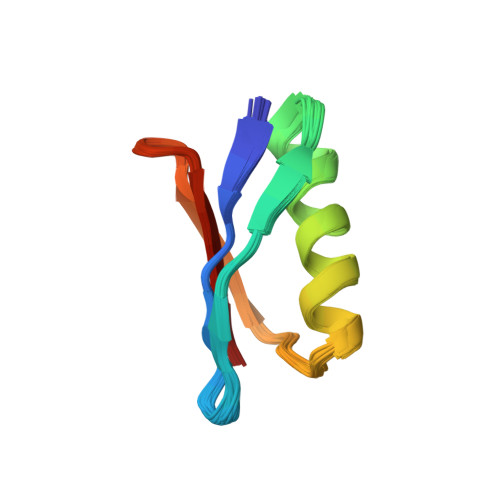
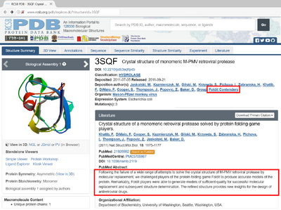
RCSB PDB - 6L2W: Crystal structure of a novel fold protein Gp72 from the  freshwater cyanophage Mic1
RCSB PDB - 6L2W: Crystal structure of a novel fold protein Gp72 from the freshwater cyanophage Mic1

Flavodoxin fold - Wikipedia

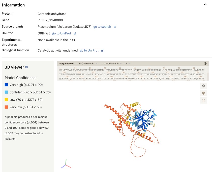
AlphaFold Protein Structure Database

Search results for PDB entry 1pvl | PDBeFold

The 3D structure of irregular β-trefoil fold proteins used in this... |  Download Scientific Diagram
The 3D structure of irregular β-trefoil fold proteins used in this... | Download Scientific Diagram
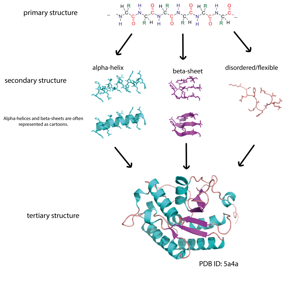
RCSB PDB - 1GB1: A NOVEL, HIGHLY STABLE FOLD OF THE IMMUNOGLOBULIN BINDING  DOMAIN OF STREPTOCOCCAL PROTEIN G
RCSB PDB - 1GB1: A NOVEL, HIGHLY STABLE FOLD OF THE IMMUNOGLOBULIN BINDING DOMAIN OF STREPTOCOCCAL PROTEIN G